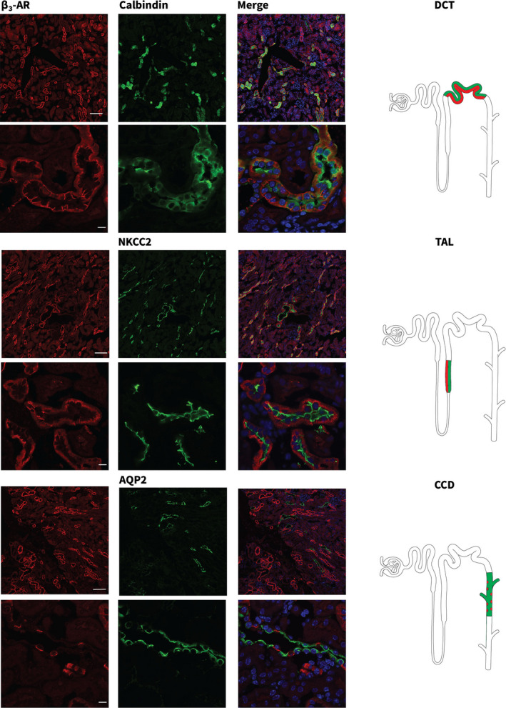
FIGURE 1.

β3‐AR localizes to multiple segments of the mouse nephron. Cryosections from wild‐type (wt) mouse kidneys were stained with anti‐β3‐AR (red) and nephron segment‐specific markers (green). Calbindin for distal convoluted tubules (DCTs), Na+K+2Cl (NKCC2) co‐transporter for the thick ascending limbs (TALs) and Aquaporin 2 (AQP2) for the cortical collecting ducts (CCDs). Confocal microscopic images were acquired at two different magnifications (Upper panels, scale bar = 100 µm; Lower panels, scale bar = 10 µm). Nuclei were stained with Hoechst Nuclear Staining (blue). Positive nephron segments are depicted in the cartoons on the right and colocalization is highlighted in red and green
