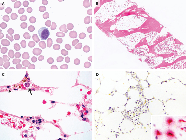
Figure 2. Peripheral-Blood and Bone Marrow Specimens.
Wright–Giemsa staining of a peripheral-blood smear (Panel A) shows a circulating plasmacytoid lymphocyte; platelets are notably absent, a finding consistent with thrombocytopenia. Hematoxylin and eosin staining of bone marrow (Panel B) shows markedly hypocellular marrow. At higher magnification (Panel C), the residual cellularity is composed mainly of lymphocytes (arrowhead) and plasma cells (arrow). In situ hybridization of bone marrow to detect severe acute respiratory syndrome coronavirus 2 (Panel D) is negative, without the red chromogen staining that indicates the presence of viral RNA; the inset shows an example of positive cellular staining in human lung tissue.

