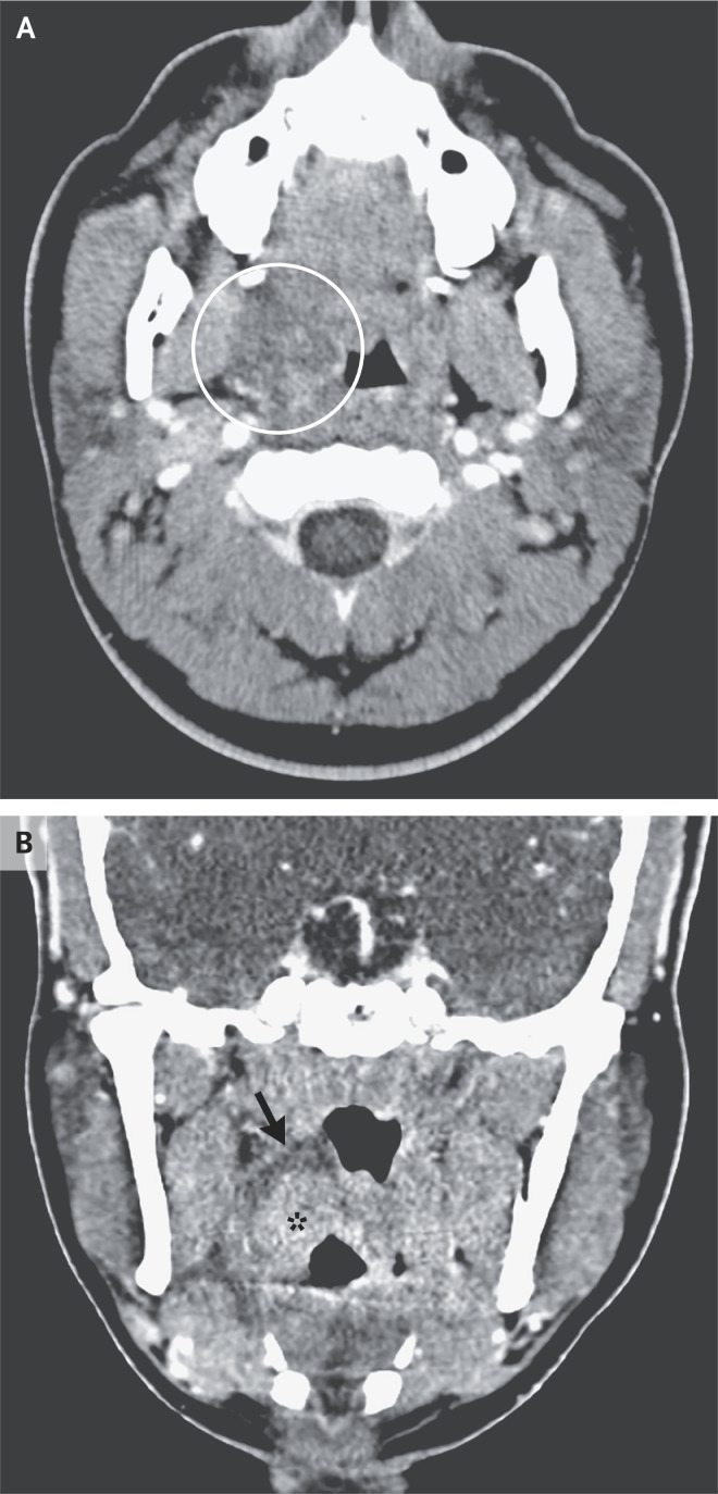
Figure 3. CT Scan of the Neck.
CT of the neck was performed after the administration of intravenous contrast material in a soft-tissue algorithm. An axial image (Panel A) shows hypoattenuating peritonsillar phlegmon (circled), which is causing partial effacement of the oropharyngeal airway. A coronal image (Panel B) shows the rim of the hypoattenuating phlegmon in the peritonsillar space (arrow) adjacent to the enlarged right palatine tonsil (asterisk). There is no evidence of a well-defined rim-enhancing abscess.

