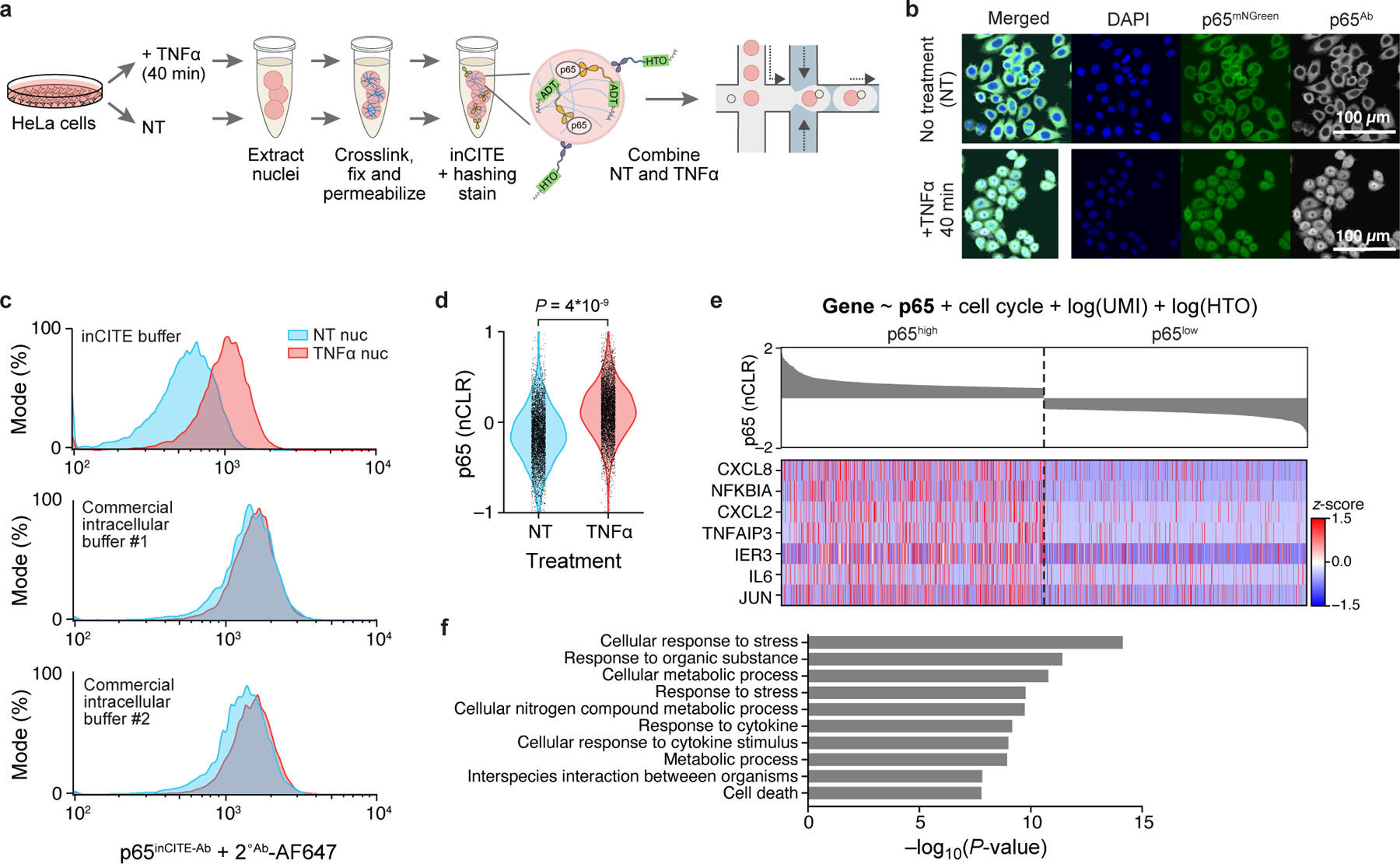
Figure 1. InCITE-seq simultaneously measures intranuclear protein and RNA levels at single nucleus resolution.

a. Overview of inCITE-seq for droplet-based profiling of nuclear proteins with nucleus hashing in HeLa cells. b. In situ fluorescent images of HeLa cells expressing a p65-mNeonGreen reporter (p65mNGreen) stained with anti-p65 antibody (p65Ab followed by Alexa Fluor 657 conjugated secondary), sampled without treatment (no treatment, “NT”; top) or 40 min after TNFα treatment (bottom); representative of 4 independently conducted experiments. Scale bar, 100μm. c. Flow cytometry of HeLa nuclei stained with p65inCITE-Ab followed by Alexa Fluor 647 secondary (x axis) sampled from NT (blue) or 40 min after TNFα treatment (red). Buffers, from top to bottom: optimized inCITE buffer with dextran sulfate, commercial buffer #1, commercial buffer #2 (Methods). d. Distribution of p65 levels (nCLRs) in NT (blue) and TNFα treated (red) nuclei profiled by inCITE-seq (P=4*10−9, two-sided Kolmogorov-Smirnov test). e. Expression (Z score, color bar) of the top 7 genes (rows) positively associated with p65 levels identified by a linear model (top, Methods) across nuclei (columns), visualized for the top decile (p65high) and bottom decile (p65low) of p65 nuclear protein levels by inCITE-seq (bar plot, top, nCLR). f. Top 10 Gene Ontology terms (y axis) significantly enriched (−log10(P-value), x axis, hypergeometric test) in 142 genes positively associated with p65 levels.
