
Extended Data Fig. 4. Impact of tissue preparation on epitope detection by antibodies.
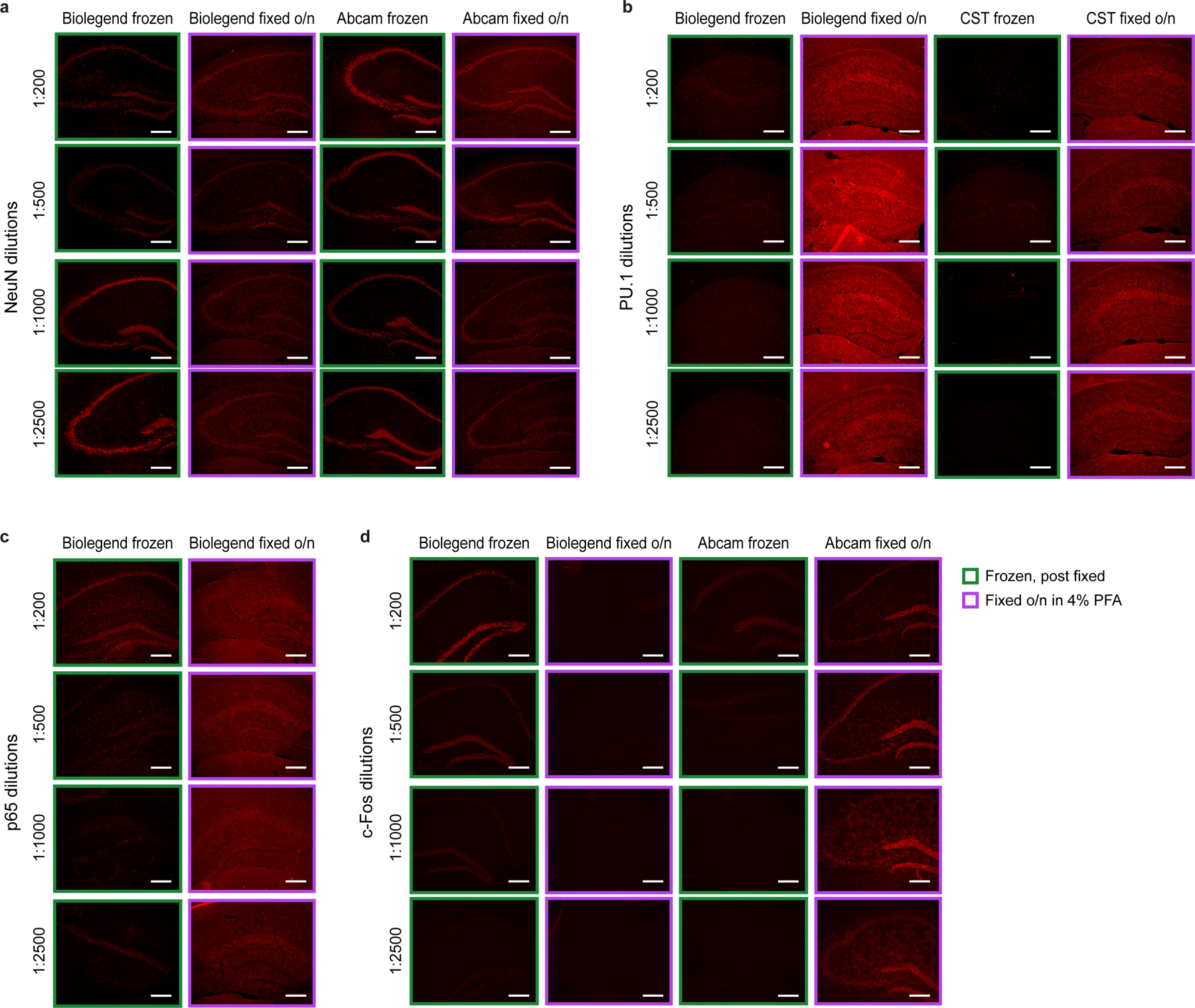
Comparing in situ immunofluorescence of antibody stains (followed by Alexa Fluor 647-conjugated secondary stain) in mouse hippocampus tissue that were immediately frozen (green box) or frozen after overnight fixation in 4% PFA (purple box, Methods) across a wide range of antibody dilutions. Images are representative of 2 independent experiments. a. NeuN in PBS. Biolegend NeuN antibody (clone 1B7) used for inCITE and Abcam NeuN antibody (clone EPR12763). b. PU.1 in PBS. Biolegend PU.1 antibody (clone 7C2C34) used for inCITE and Cell Signaling Technology PU.1 antibody (clone 9G7). c. p65 in KA. Biolegend p65 antibody (clone Poly6226) used for inCITE. d. c-Fos in KA treated mice. Biolegend c-Fos antibody (clone Poly6414) used for inCITE and Abcam c-Fos antibody (ab190289). Scale bars, 100μm.