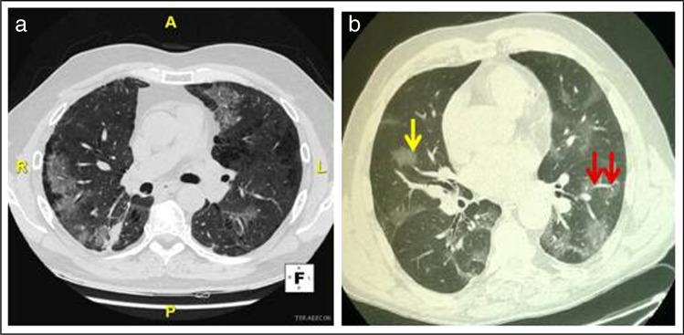
Fig. 1.
CT scans of pneumonia due to a immune checkpoint inhibitor therapy or b COVID-19: a Axial lung image (without intravenous contrast) of an immune checkpoint inhibitor-treated 63-year-old man with metastatic non-small cell lung cancer, showing ground glass opacities, with nonrounded morphology and no specific distribution, associated to a right small area of consolidation. b Axial lung image (without intravenous contrast) of a 64-year-old man with COVID-19 showing bilateral ground glass opacities someone with rounded morphology (yellow arrow) and interlobular septal thickening, predominantly located in the peripheral, near the fissures and posterior part of the lungs; vascular dilation is also seen (red arrows)

