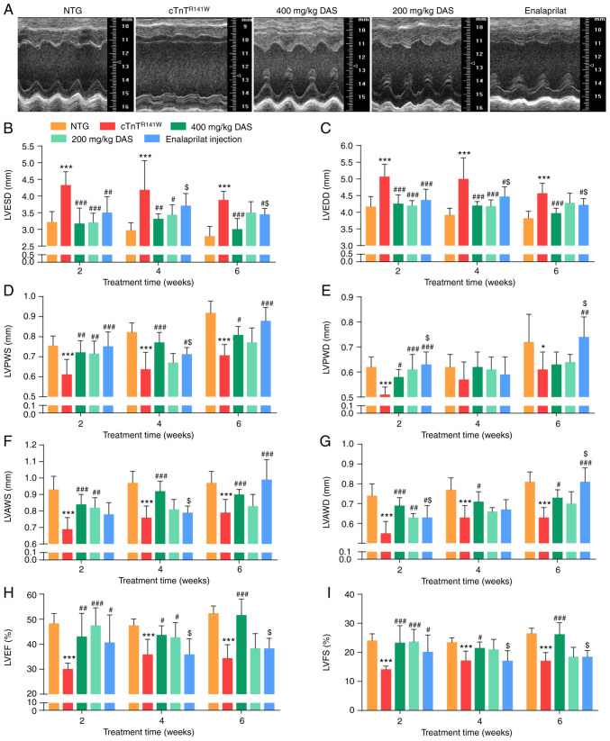
Figure 1.
Echocardiographic analysis of cardiac morphology and function. Mice in the five groups were analyzed after 6 weeks of treatment: NTG (wild-type control), cTnTR141W (placebo control), treatment with 400 mg/kg DAS, treatment with 200 mg/kg DAS and treatment with enalaprilat (commercially available drug control). (A) Representative M-mode echocardiographic images of the LV long axis. (B) LVESD. (C) LVEDD. (D) LVPWS. (E) LVPWD. (F) LVAWS. (G) LVAWD. (H) LVEF. (I) LVFS. n=7. *P<0.05, ***P<0.001 vs. NTG group; #P<0.05, ##P<0.01, ###P<0.001 vs. cTnTR141W group; $P<0.05 vs. 400 mg/kg DAS group. NTG, non-transgenic; LV, left ventricular; LVESD, left ventricular end-systole diameter; LVEDD, left ventricular end-diastole diameter; LVPWS, left ventricular ventricle posterior wall at end systole; LVPWD, left ventricular posterior wall at end diastole; LVAWS, left ventricular anterior wall at end systole; LVAWD, left ventricular anterior wall at end diastole; LVEF, left ventricular ejection fraction; LVFS, left ventricular fractional shortening; DAS, diallyl sulfide.

