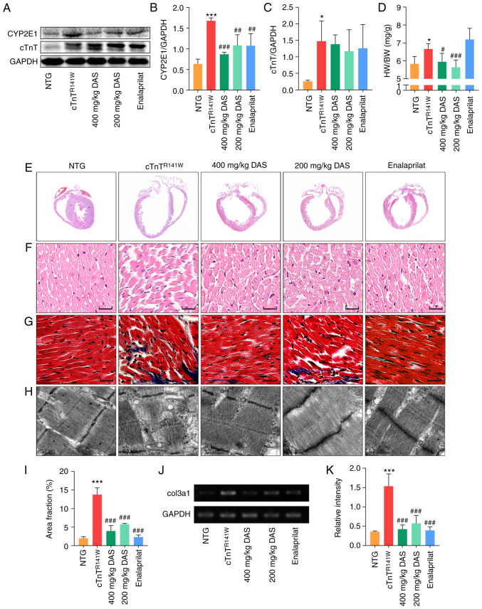
Figure 2.
Pathological histology observation. (A) CYP2E1 and cTnT expression in the heart tissues of mice in the NTG, cTnTR141W, 400, 200 mg/kg DAS and enalaprilat groups was detected via western blotting. (B) CYP2E1 and (C) cTnT were semi-quantitatively analyzed, using GAPDH for normalization (n=3). (D) Ratio of HW to BW (n=6). (E) H&E staining patterns of whole-heart longitudinal sections. (F) Magnification of H&E-stained sections of the left ventricle (magnification, ×400; scale bar, 20 µm). (G) Magnification of Masson's trichrome-stained left ventricle sections (magnification, ×400, scale bar, 20 µm). Myocytes are stained red; collagenous tissue is stained blue. (H) Ultrastructure observation via transmission electron microscopy (scale bar, 0.5 µm). (I) Quantitative analysis of Masson staining (n=3). (J) Col3α1 expression was detected and (K) analyzed (n=3). *P<0.05, ***P<0.001 vs. NTG group; #P<0.05, ##P<0.01, ###P<0.001 vs. cTnTR141W mice. cTnT, cardiac troponin T; HW, heart weight; BW, body weight; Col3α1, procollagen type III α1; NTG, non-transgenic; DAS, diallyl sulfide; CYP2E1, cytochrome P450 family 2 subfamily E member 1.

