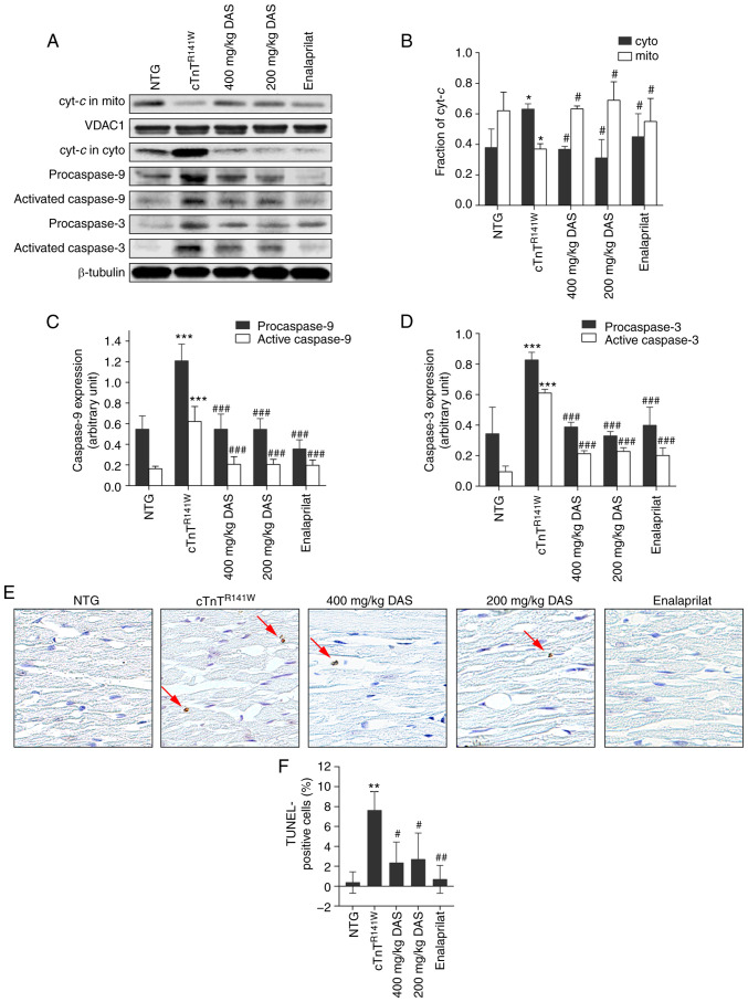
Figure 4.
Analysis of the mitochondria-dependent apoptosis pathways. (A) Cyt-c release and activation of caspase 9 and caspase 3 in the heart tissues of mice in the NTG, cTnTR141W, 400, 200 mg/kg DAS and enalaprilat groups were detected via western blotting. (B) Cyt-c in the cyto and mito was semi-quantitatively analyzed using β-tubulin or VDAC1 for normalization. (C) Procaspase 9 and (D) procaspase 3 and active caspases 3 and 9 were semi-quantitatively analyzed using β-tubulin for normalization (n=3). (E) Cardiac myocyte apoptosis was detected using a TUNEL assay, and the arrows indicate TUNEL-positive cells (magnification, ×400). (F) Number of positive cells was counted, and the proportion of apoptotic cells among the total cells in each image was calculated (n=3). *P<0.05, **P<0.01, ***P<0.001 vs. NTG group; #P<0.05, ##P<0.01, ###P <0.001 vs. cTnTR141W mice. Cyt-c, Cytochrome c; cyto, cytoplasm; mito, mitochondria; VDAC1, voltage dependent anion channel 1; NTG, non-transgenic; DAS, diallyl sulfide.

