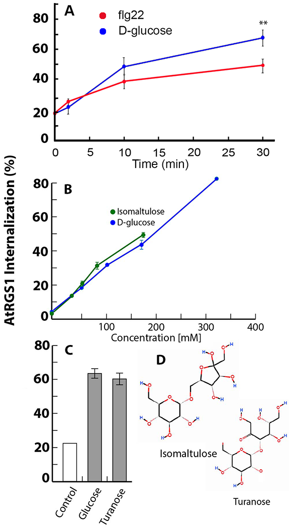
Fig. 1. An AtRGS1 complex perceives extracellular flg22 and glucose or glucose metabolite.

(A) Endocytosis of AtRGS1-YFP induced by flg22 (red) or D-glucose (blue). Values represent the amount of AtRGS1 that is internal to the cell as a percent of the total YFP fluorescence. The type of raw data used to generate these values is shown in Supplemental Fig. S1C. **, P < 0.01. N=4-24 biological replicates per group. (B) Endocytosis of AtRGS1-YFP induced by isomaltulose (green) or d-glucose (blue). Supporting data is in Fig. S1C to E,N=5 biological replicates per group (C) Endocytosis of AtRGS1-YFP induced by turanose or d-glucose. The purity of turanose was >98%. N=12 biological replicates per group. (D) Structures of isomaltulose and turanose, which both contain a glucose ring moiety.
