Figure 1.

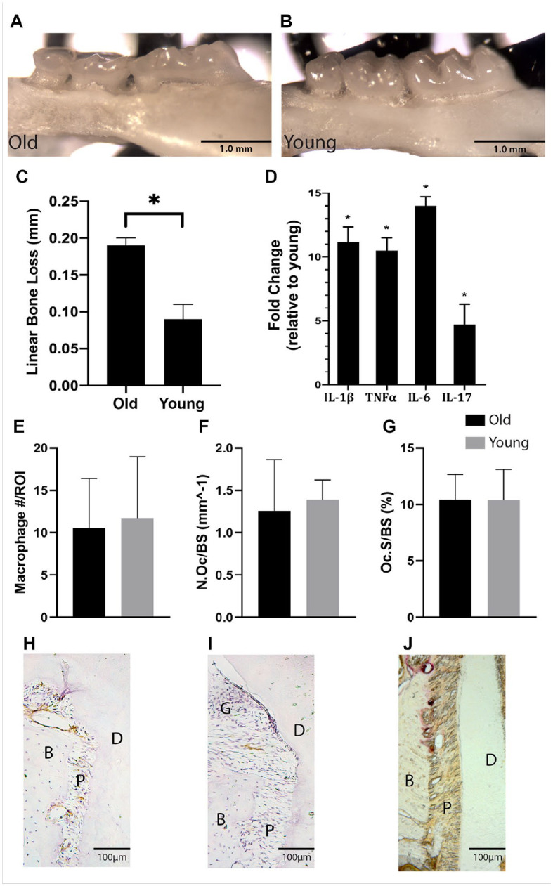
Old mice demonstrate an aged periodontal phenotype. The maxillae of control old (24 mo) and young (3 mo) male mice (C57BL/6J) were collected and analyzed (A, B). Significantly increased linear bone loss, measured from the cementoenamel junction to the alveolar bone crest, was demonstrated in old mice (C). The increased bone loss was associated with a significant increase in proinflammatory cytokine expression within the gingiva as measured by quantitative real-time polymerase chain reaction (D). Frozen histological sections were stained for F4/80 and tartrate-resistant acid phosphatase (TRAP), and F4/80+ macrophages, TRAP+ cell surface/bone surface, and TRAP+ osteoclast number per bone surface were quantified. Macrophage and osteoclast quantity in the periosteum were similar in old and young mice (E–G). Histomorphometry demonstrates the presence of F4/80+ macrophages (DAB, brown) (H, I) and TRAP+ osteoclasts (red) (J) within the periodontium in controls. B, bone; D, dentin; G, gingival tissue; P, periodontal ligament. Data presented as mean ± SD. *P < 0.05.
