Figure 3.

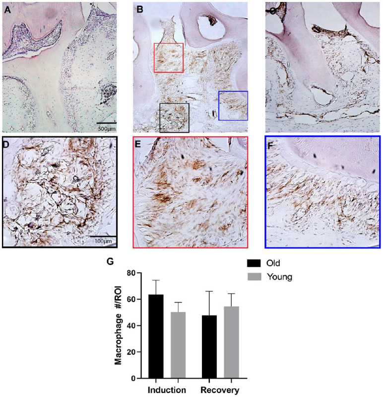
Macrophage quantity within the periodontium is similar in young and old mice during disease induction and recovery. Immunohistochemistry staining for F4/80+ macrophages was performed on frozen histological sections of the periodontium of old and young mice. F4/80+ macrophages (DAB, brown) were present in healthy controls (A), after 7 d of disease inductions (B), and after 7 d of disease recovery (C). Macrophages were quantified within 3 distinct regions of interest (ROIs) per histological section. ROIs were identified within an osseous region (D), gingival tissue region (E), and periodontal ligament (PDL) region (F). Macrophage quantity within the periodontium during disease induction and recovery was similar in old and young mice (n = 6/group) (G). Data presented as mean ± SD.
