
Fig. 7.
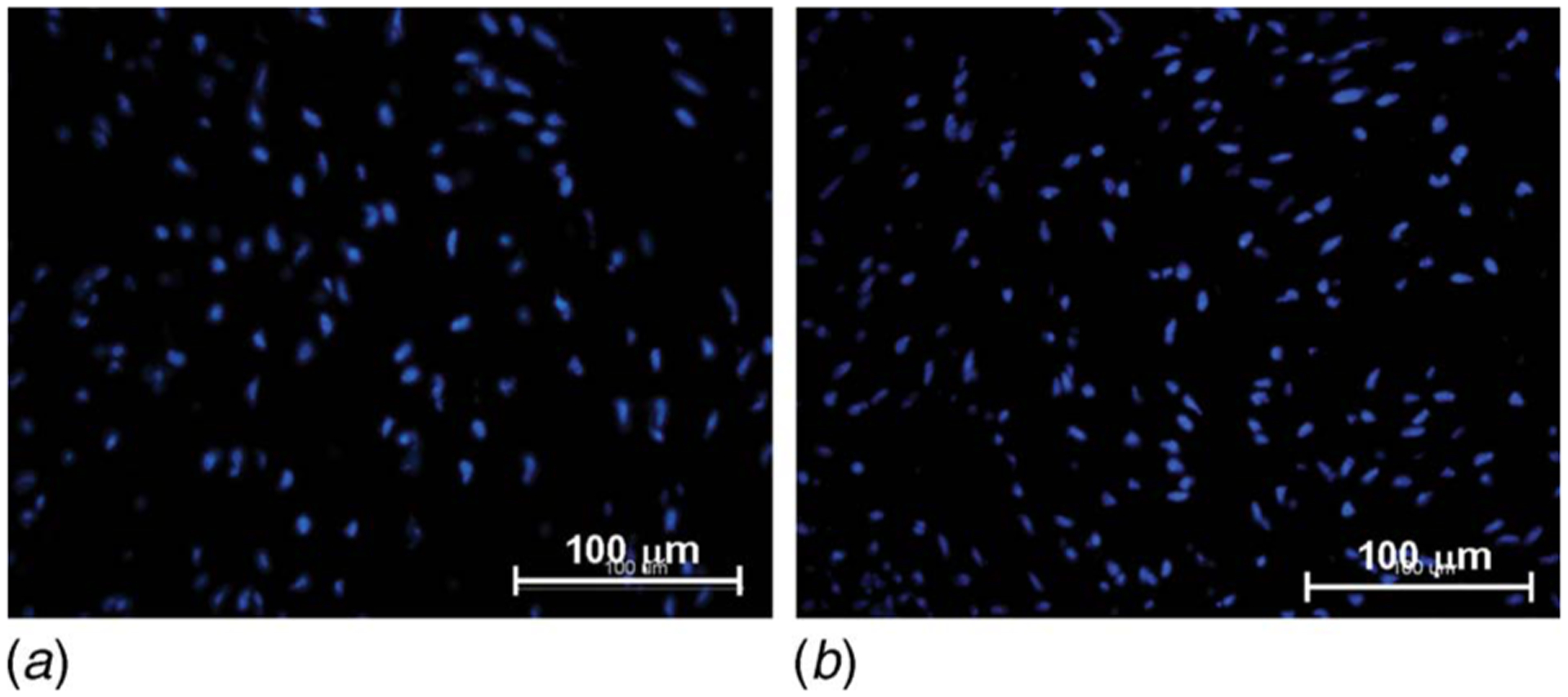
DAPI stain on (a) aortic valve leaflet exposed to physiologic ventricular shear stress conditions for 120 h and (b) fresh control (nuclei stained in blue)
Official websites use .gov
A
.gov website belongs to an official
government organization in the United States.
Secure .gov websites use HTTPS
A lock (
) or https:// means you've safely
connected to the .gov website. Share sensitive
information only on official, secure websites.
DAPI stain on (a) aortic valve leaflet exposed to physiologic ventricular shear stress conditions for 120 h and (b) fresh control (nuclei stained in blue)