
Figure 1.
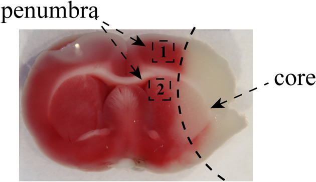
The images of the penumbra after MCAO. The images showed that the black zone marked as “1” represented the selected cortex region near the infarct core and the area labeled as “2” was marginal striatum around the infarct core. We used the brain tissue from penumbra to perform qRT-PCR, Luminex multiplex assay, and Western blotting. The area of penumbra in the cortex and striatum for immunofluorescence staining was chosen as shown.