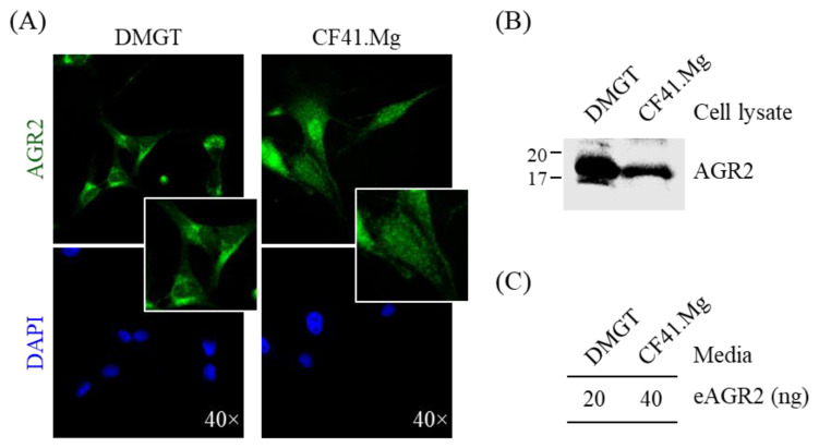
Figure 3.
Detection of AGR2 expression in canine MMT cells and conditioned media. (A) Intracellular localization of AGR2 in canine MMT cell lines, DMGT, and CF41.Mg. AGR2 expression was verified by immunofluorescence staining using an AGR2-specific antibody. DAPI staining indicated the nuclei. Magnification, ×40. (B) Detection of AGR2 in cell lysates of canine MMT cell lines. Cell lysates (30 µg proteins per sample) were analyzed by immunoblotting with an AGR2-specific antibody. (C) Detection of extracellular AGR2 (eAGR2) in serum-free conditioned media of canine MMT cells. Cells were grown in serum-free media for one day, and the conditioned media were harvested and subsequently concentrated. The resulting conditioned media (50 µg proteins per sample) were analyzed using a competitive ELISA established for eAGR2 detection.

