Figure 2.
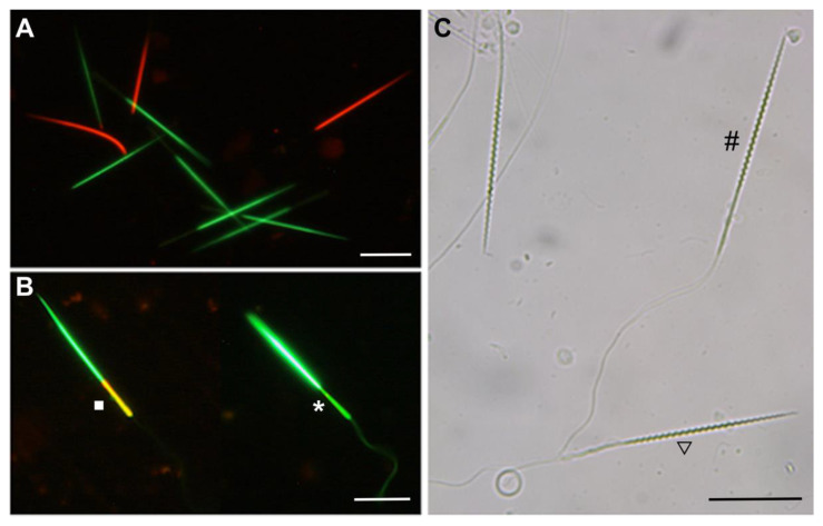
Representative images of small-spotted catshark (Scyliorhinus canicula) sperm cell quality. Scale bar, 20 μm. (A) Epifluorescence micrographs of sperm cells stained with SYBR-14 (green) and propidium iodide (red) at 200× magnification. Green fluorescence shows live sperm, and red fluorescent indicates dead sperm. (B) Epifluorescence micrographs of sperm cells stained with JC-1 at 200× magnification. The dye changes emission wavelength depending on membrane potential by a shift from orange, showing high potential (square), to green color, showing low potential (asterisk). (C) Phase-contrast image displaying tail swelling to assess the functional integrity of membrane by hypoosmotic swelling test at 200× magnification. Coiled tail spermatozoa were identified as having a functional intact plasma membrane (triangle); normal tail spermatozoa were identified as having a nonfunctional intact plasma membrane (octothorpe).

