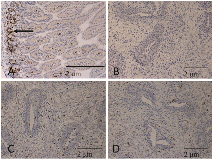
Figure 3.
Tissues immunostained for KI67 (brown) and counterstained with hematoxylin. (A) Jejunum was collected from gilts and used as a positive control for KI67 labeling of proliferating cell populations in tissues. Arrow indicates crypt region of villi. Immunostaining of KI67 shows that epithelial cells within the crypt region of the villi were immunostained, whereas those lining the villi of the jejunum were mostly lacking positive cells. (B) Negative control mammary tissue was incubated with rabbit IgG rather than primary antibody and show a lack of brown nuclei. Mammary tissue from (C) COL10 and (D) COL20 gilts immunostained with KI67 have positively stained cells in the epithelial and stromal components of the parenchymal compartment. Images were captured at 200×.

