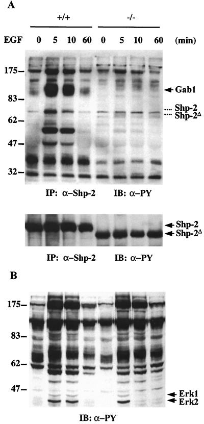
FIG. 3.
EGF-induced association of Shp-2 with tyrosine-phosphorylated proteins. (A) Shp-2 protein was immunoprecipitated (IP) from wild-type (+/+) or mutant (−/−) cell lysates, resolved by SDS-PAGE, and immunoblotted (IB) with anti-PY antibody. The same membrane was then stripped and reprobed with an anti-Shp-2 antibody raised against the C-terminal region of Shp-2. The corresponding positions of wild-type and mutant Shp-2 bands were indicated with dashed lines in the upper panel. (B) Equal amounts of whole cell lysates from control or EGF-stimulated cells were resolved by SDS-PAGE, transferred to a nitrocellulose membrane, and blotted with an anti-PY antibody.

