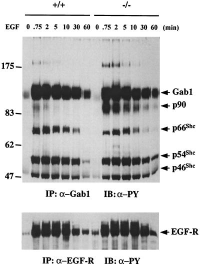
FIG. 7.
Functional interaction between Gab1 and Shp-2. Total cell lysates (1 mg of total proteins) from Shp-2+/+ or Shp-2−/− cells stimulated by 100 ng of EGF per ml for the indicated times were mixed with 2 μl of anti-Gab1 antibody and protein A-Sepharose 4B beads for immunoprecipitation (IP). The precipitates were then immunoblotted (IB) with anti-PY antibody to assess the tyrosine phosphorylation levels of Gab1 as well as Gab1-associated phosphoproteins. In the lower panel, the EGF-R was immunoprecipitated from the same cell lysates and immunoblotted with anti-PY antibody.

