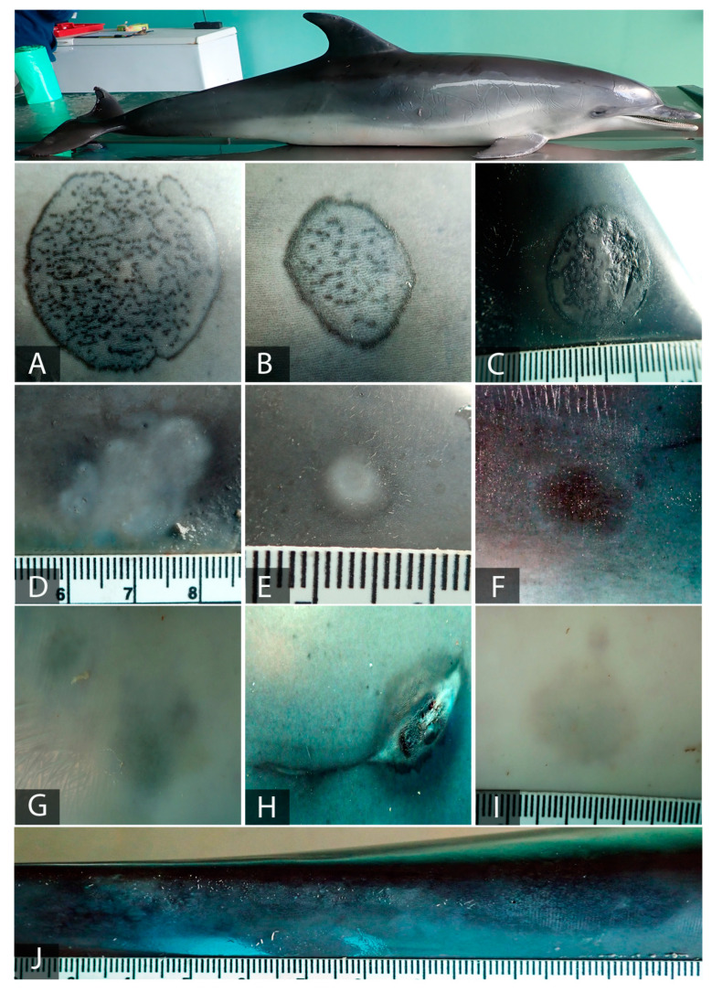
Figure 3.
Gross lesions compatible with CePV in Atlantic spotted dolphin, Case 2. Right lateral view. (A) Ring lesion with a black edge and stippled pattern center (3 × 2.3 cm) on the right side of the melon. (B) Ring lesion with a black edge and stippled pattern center (1 × 0.7 cm) located on the right side of the melon. (C) Oval lesion presenting both margin and inner ping-hole pattern slightly raised with half of the center blistered (1.8 × 1.3 cm), located on the right side of the dorsal fin. (D) Irregular pale and coalesced wound with a barely visible dark edge (2.3 × 1.2 cm) located on the right dorsolateral superior hemimaxilla. (E) An oval lesion with a pale center and blurred margin (0.6 × 0.3 cm) situated on the right dorsal part of the tip. (F) Oval dark lesion with pale margin (1.5 × 1 cm) located on the right side of the animal. (G) A blurred and irregular grey lesion on the rostroventral part. (H) An oval dark lesion with a pale, raised, and irregular center (1.6 × 1.3 cm) on the right lateral side. (I) A blurred hardly visible grey lesion (1.8 × 1.5 cm) on the ventral part of the animal. (J) A large and irregular dark lesion with a greyish pin-hole pattern across the entire center located on the dorsal part of the peduncle.

