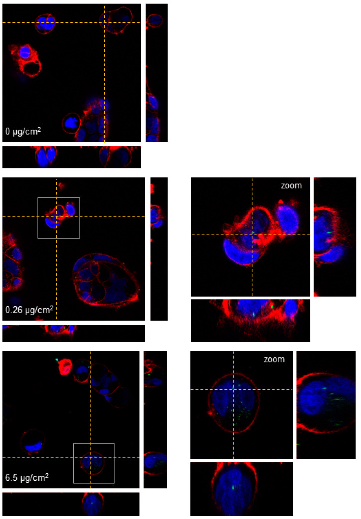
Figure 3.
Three-dimensional confocal microscopy images of undifferentiated Caco-2 cells after a 24 h exposure to y-PSNPs. Nuclei are stained in blue, cell membranes in red, and nanoparticles are depicted in green. Dotted lines point out the plane from where the orthogonal views are projected. Images on the left correspond to cells exposed to the different concentrations of y-PSNPs indicated while images on the right correspond to the zoomed area highlighted by a grey square.

