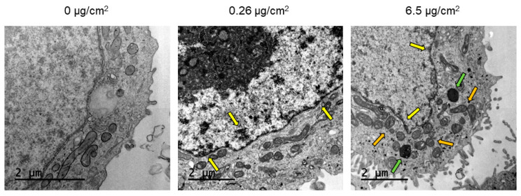
Figure 4.
TEM images of Caco-2 cells after 24 h of exposure to increasing concentrations of PSNPs. Dark and electron-dense formations observed in the perinuclear region are pointed out with yellow arrows, while PSNPs accumulations in vacuoles and lysosomes are indicated with green arrows. Orange arrows point out the induction of mitochondrial cristae swelling.

