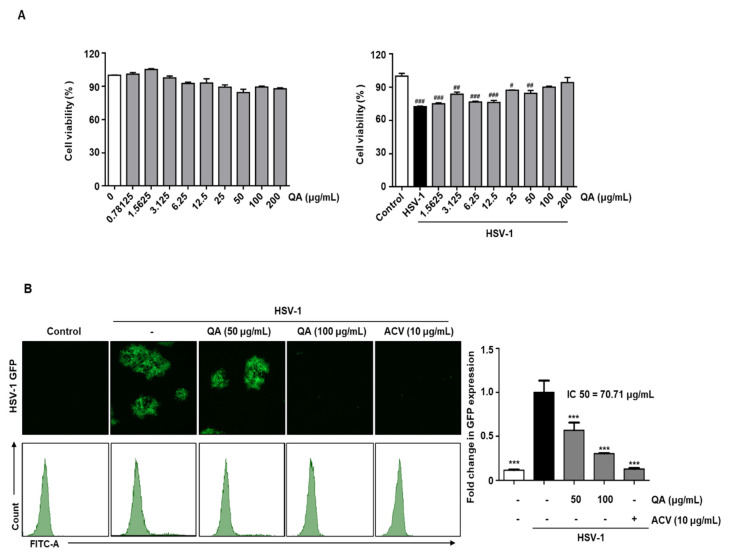
Figure 3.
QA exhibits antiviral effects upon infection with HSV-1 in SK-N-SH cells. (A) SK-N-SH cells were treated with QA at the indicated concentrations for 48 h (left). SK-N-SH cells were infected with HSV-1 strains (MOI = 0.01) for 2 h and then treated with QA at the indicated concentrations for 48 h (right). Cell viability was measured by CCK-8 assay. (B) SK-N-SH cells were infected with HSV GFP (MOI = 2) for 2 h and then treated with QA (50 and 100 μg/mL) for 48 h. HSV GFP expression levels were analyzed by fluorescence microscopy (left) and flow cytometry (right). The data are representative of three independent experiments and quantified as mean values ± SEM. One-way ANOVA with Tukey’s post hoc test; # p < 0.05, ## p < 0.01, ### p < 0.001, compared with the control. *** p < 0.001, compared with the HSV-1treatment. IC50; The half maximal inhibitory concentration.

