Figure 5.
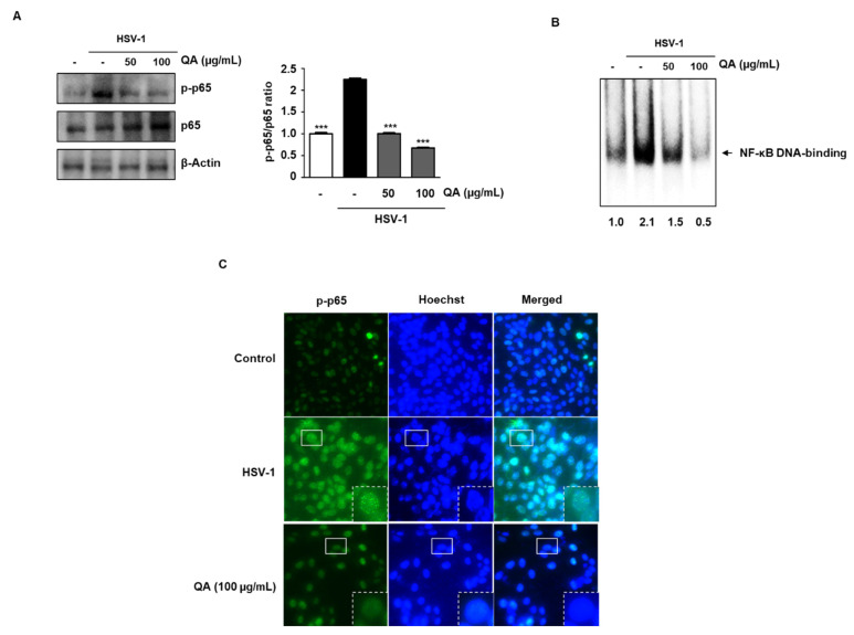
QA inhibits NF-κB phosphorylation by HSV-1 infection in SK-N-SH cells. Cells were infected with HSV-1 (MOI = 0.01) for 2 h and then treated with QA at 50 and 100 μg/mL for 48 h. (A) Whole cell extracts were subjected to western blot analysis for p-p65 and p65. β-actin was used as an internal control. (B) QA suppresses constitutive activation of NF-κB by HSV-1 (MOI = 0.01) infection in SK-N-SH cells. Cells were infected with HSV-1 and then incubated for 48 h with the indicated concentrations of QA. Then, nuclei were extracted from SK-N-SH cells, and NF-κB activation was analyzed by EMSA. (C) The intracellular distribution of p-p65 was analyzed by an immunofluorescence assay. The third panel displays the merged images of the first and second panels. The data are representative of three independent experiments and quantified as mean values ± SEM. One-way ANOVA with Tukey’s post hoc test; *** p < 0.001, compared with the HSV-1treatment.

