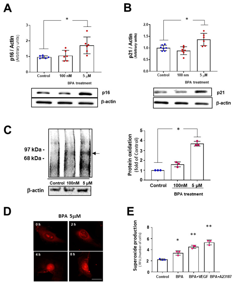
Figure 2.
BPA induces senescent protein markers p21 and p16 and increases oxidative stress. Western blotting analysis of MAEC treated with 100 nM and 5 µM BPA for five days using antibodies against (A) p21 and (B) p16. β-actin was used as a loading control. (n = 6). Results are given as mean ± SD, p was determined by a Kruskal–Wallis test for the comparison between control and BPA-treated cells.* p < 0.05 ); (C) The levels of reactive oxygen species induced in MAEC by BPA were analyzed by oxyblot assay in 100 nM and 5 µM BPA-treated MAEC for 24 h to detect carbonyl groups in proteins as a marker of protein oxidation. The densities of the proteins between 97 and 68 KDa were normalized using the expression of β-actin. (n = 3 with duplicate for each condition).* p < 0.001 using Kruskal–Wallis test for the comparison between control and BPA-treated cells; (D) Immunofluorescence (IF) of superoxide production in MAEC in response to 5 µM BPA for 0–8 h, using the fluorescence probe dihydroetidium (DHE). After 4 h, fluorescence can be observed at the cell nucleus. Scale bar: 10 µm; (E) Quantification of superoxide in MAEC upon 5 µM BPA treatment for 24 h, BPA+VEGF and BPA+ A23187 and vehicle (control). (n = 3, per duplicate). Results are given as mean ± SD, p was determined by a Kruskal–Wallis test, * p < 0.001 vs. Control and ** p < 0.001 vs. BPA.

