Figure 1.
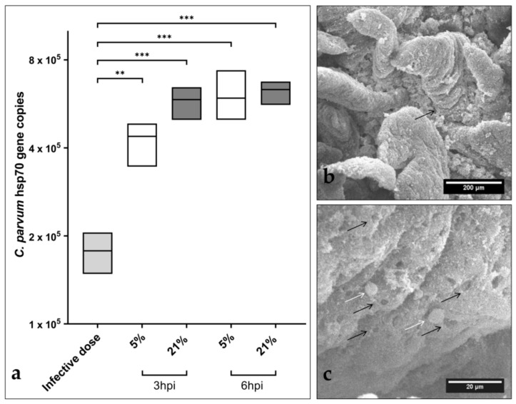
Early Cryptosporidium parvum development in bovine small intestinal (BSI) explants under both physioxic (5% O2) and hyperoxic (21% O2) conditions. A significant increase in parasite replication was detected in BSI explants (n = 3) via C. parvum hsp 70 gene-specific qPCR analyses, and by comparing 3 and 6 hpi with the initial sporozoite infection dose (i.d.), boxplots represent mean ± SD (a). SEM analysis of BSI explants confirmed parasite replication by revealing C. parvum-infected villi (black arrow) (b). Interestingly, C. parvum-infected BSI explants presented typical C. parvum-induced hole-like lesions in epithelial cells (black arrows), along with development of trophozoite-like stages (white arrows), which were detected as early as 3 hpi (c). Statistical significance (** p < 0.01, *** p < 0.001) was determined by Kruskal–Wallis test followed by Dunn’s multiple comparison test comparing infected BSI explants with initial sporozoite numbers used for infection (infection doses, n = 3). qPCR- and SEM-based analyses were performed in duplicate and triplicate, respectively.

