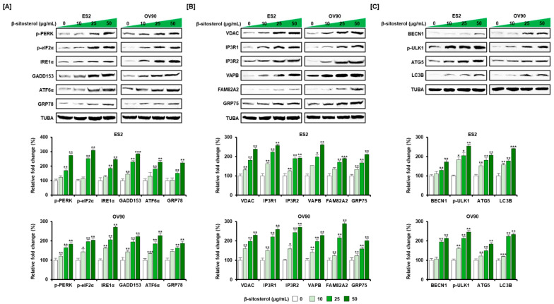
Figure 4.
Increased expression of ER stress sensors and ER-mitochondrial axis and autophagy proteins by β-sitosterol in ovarian cancer cells. Western blot bands of UPR proteins (A) including protein kinase R (PKR)-like endoplasmic reticulum kinase (PERK), eukaryotic translation initiation factor 2α (eIF2α), inositol-requiring enzyme-1α (IRE1α), growth arrest and DNA damage 153 (GADD153), activating transcription factor 6α (ATF6α), 78 kDa glucose-regulated protein (GRP78), ER-mitochondrial axis proteins (B) voltage-dependent anion channel (VDAC), IP3 Receptor 1 (IP3R1), IP3 Receptor 2 (IP3R2), vesicle-associated membrane protein (VAPB), regulator of microtubule dynamics 3, RMDN3 (FAM82A2), glucose-regulated protein 75 (GRP75), and autophagy proteins (C) beclin 1 (BECN1), UNC-51-like kinase 1 (ULK1), autophagy-related 5 (ATG5), autophagy marker Light Chain 3 B (LC3B) after β-sitosterol (0, 10, 25, and 50 µg/mL) treatment. Alpha-tubulin (TUBA), used as a control, is shown at the bottom of each set. The asterisks indicate significant differences between treated and control cells (*** p < 0.001, ** p < 0.01, and * p < 0.05).

