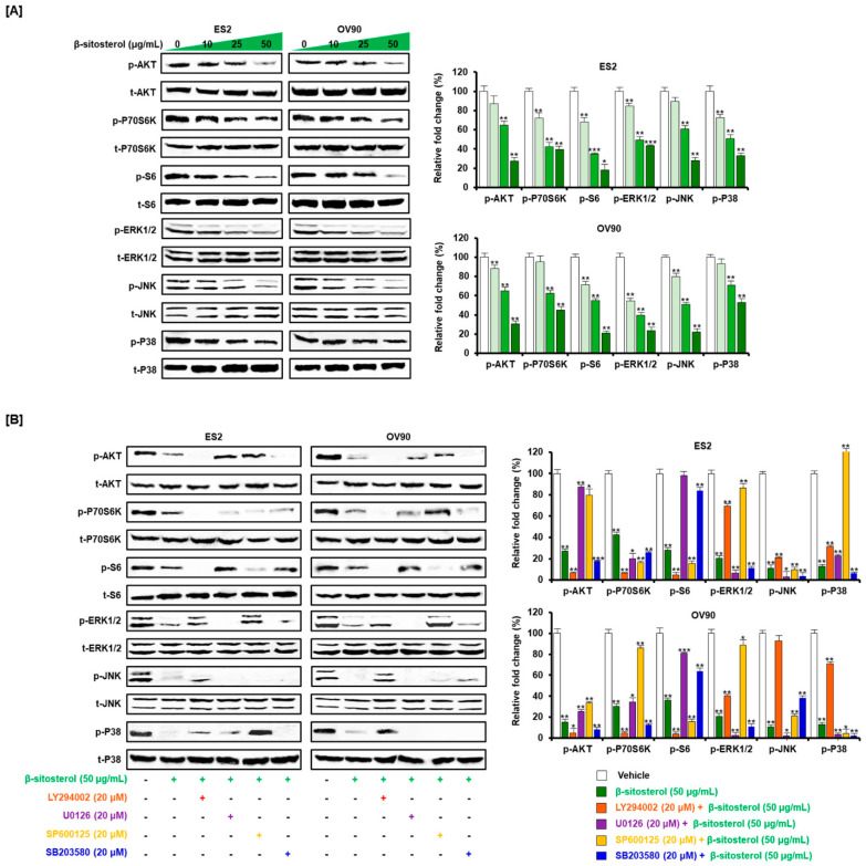
Figure 6.
Change of cell growth-related signals by β-sitosterol in the two cell lines. (A) Western blots show phosphorylation changes in the PI3K pathway including protein kinase B (AKT), P70S6 kinase (P70S6K), S6 and in the MAPK pathway including extracellular signal-regulated kinase 1/2 (ERK1/2), c-Jun N-terminal kinase (JNK), P38, in ovarian cancer cells after β-sitosterol (0, 10, 25, and 50 µg/mL) treatment. (B) Western blots show changes in the phosphorylation levels of proteins in the PI3K and MAPK signaling pathways in ovarian cancer cells following co-treatment with β-sitosterol and each inhibitor. The asterisks indicate significant differences between treated and control cells (*** p < 0.001, ** p < 0.01, and * p < 0.05).

