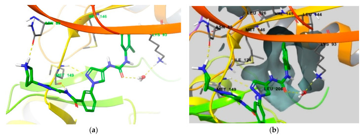
Figure 9.
(a) Crystal structure of JNK3 bound to compound 1 (PDB ID: 4WHZ [22]). (b) Protein surface of the hydrophobic pocket is shown in the same JNK3 co-crystal. Residues that interact with compound 1 and comprise selectivity pocket are emphasized in the thin tube.

