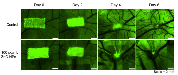
Figure 4.
Representative Images of the Development of the Chorioallantoic Membrane treated with ZnO NPs Applied onto a Gelatin Sponge. The vasculature of the chorioallantoic membrane developed rapidly from EDD 8 (day 0) to EDD 14 (day 6) (first row). Treatment with 100 µg/mL ZnO NPs onto an autofluorescent gelatin sponge (in green) did not influence the development (second row). The gelatin sponge is gradually degraded by the highly vascularized CAM over time. There were no significant negative tissue reactions observed following treatment with ZnO NPs.

