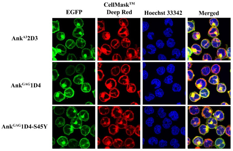
Figure 2.
Subcellular localization of ankyrins in SupT1 cells. Ankyrin-EGFP expressing SupT1 cells were stained with plasma membrane dye, CellMaskTM Deep Red. Nuclei were stained with Hoechst 33342. Confocal imaging was done at 60× magnification using Nikon C2 plus confocal fluorescence microscopy. Green represents EGFP-tagging ankyrins. Blue indicates nucleus, and red shows the plasma membrane of SupT1 cells. AnkA32D3, AnkGAG1D4, and AnkGAG1D4-S45Y refer to SupT1 cells expressing AnkA32D3, AnkGAG1D4, and AnkGAG1D4-S45Y, respectively.

