Figure 8.
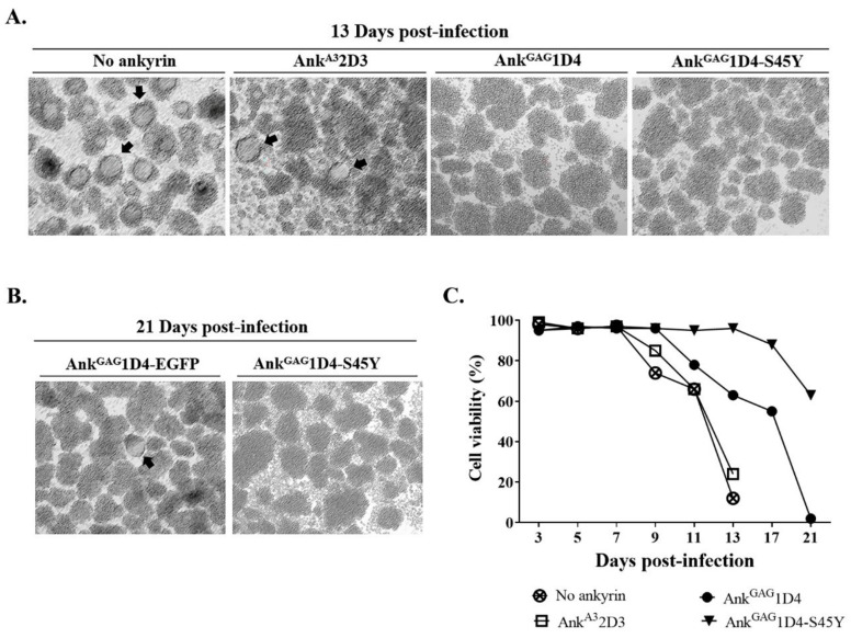
Cell morphology and cell viability of HIV-1 NL4-3 MIRCAI201V infected SupT1 stable cells. SupT1cells and ankyrin-expressing SupT1 cells were infected with 10 MOI of HIV-1 MIRCAI201V virus. After infection, cells were subcultured every 2 days. (A) Syncytium cells and cell morphology were observed under microscopy. Cell imaging was done at 10× magnification using Axio Vert.A1. (B) Cell morphology of infected SupT1/Myr (+) AnkGAG1D4-EGFP and SupT1/Myr (+) AnkGAG1D4-S45Y-EGFP was continuously observed until 21 days post-infection. Arrows point to syncytium cells. (C) Cell viability of infected cells was determined using Trypan blue exclusion method. No ankyrin, AnkA32D3, AnkGAG1D4, and AnkGAG1D4-S45Y represent SupT1 cell control, SupT1 cells expressing Myr (+) AnkA32D3-EGFP, Myr (+) AnkGAG1D4-EGFP, and Myr (+) AnkGAG1D4-S45Y-EGFP, respectively.

