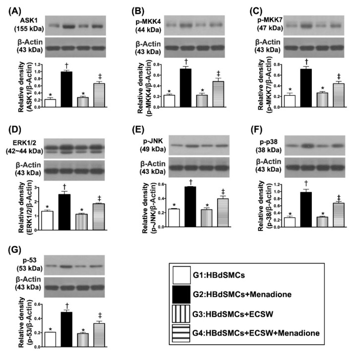
Figure 3.
ECSW therapy regulated the cell-stress signaling in RBdSMCs. (A) Protein expression of Apoptosis signal-regulating kinase 1 (ASK1), * vs. other groups with different symbols (†, ‡), p < 0.001. (B) Protein expression of phosphorylated Mitogen-activated protein kinase kinase 4 (p-MKK4), * vs. other groups with different symbols (†, ‡), p < 0.001. (C) Protein expression of p-MKK7, * vs. other groups with different symbols (†, ‡), p < 0.001. (D) Protein expression of ERK1/2, * vs. other groups with different symbols (†, ‡), p < 0.001. (E) Protein expression of phosphorylated c-Jun N-terminal kinases (p-JNK), * vs. other groups with different symbols (†, ‡), p < 0.001. (F) Protein expression of p-p38, * vs. other groups with different symbols (†, ‡), p < 0.001. (G) Protein expression of p-53, * vs. other groups with different symbols (†, ‡), p < 0.001. All statistical analyses were performed by one-way ANOVA, followed by Bonferroni multiple comparison post hoc test (n = 6 for each group). Symbols (*, †, ‡) indicate significance (at 0.05 level). ECSW = extracorporeal shock wave; RBdSMCs = rat bladder smooth muscle cells.

