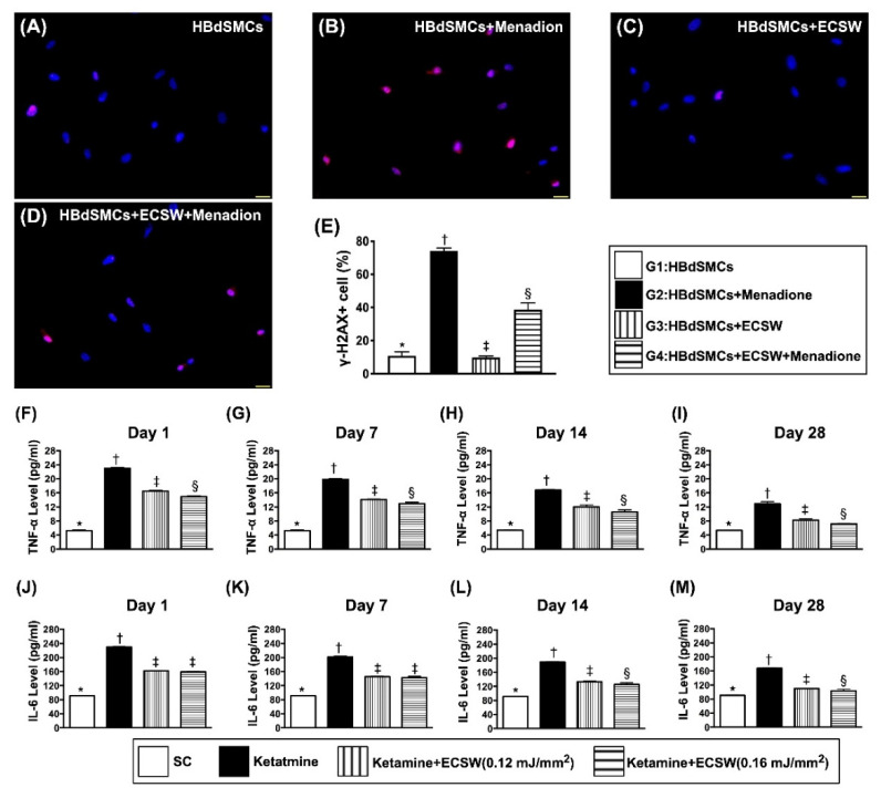
Figure 4.
ECSW therapy attenuated menadione-induced DNA damage in RBdSMCs and time courses of urinary level of inflammatory biomarkers. (A–D) Illustrating the immunofluorescent microscopic finding (400×) for identification of positively stained γ-H2AX cells (pink color). (E) Analytical result of number of γ-H2AX+ cells, * vs. other groups with different symbols (†, ‡, §), p < 0.0001. Scale bar in right lower corner represents 20 µm. (F–I) The urine levels of tumor necrosis factor (TNF)-α by days 1 (F), 7 (G), 14 (H) and 28 (I), respectively, analytical result of TNF-α in urine, * vs. other groups with different symbols (†, ‡, §), p < 0.0001. (J–M) The urine levels of interleukin (IL)-6 by days 1 (J), 7 (K), 14 (L) and 28 (M), respectively, analytical result of interleukin (IL)-6 in urine, analytical result of IL-6 in urine, for days 1 and 7, * vs. other groups with different symbols (†, ‡), p < 0.0001; for days 14 and 28, * vs. other groups with different symbols (†, ‡, §), p < 0.0001. All statistical analyses were performed by one-way ANOVA, followed by Bonferroni multiple comparison post hoc test (n = 6 for each group). Symbols (*, †, ‡) indicate significance (at 0.05 level). ECSW = extracorporeal shock wave; RBdSMCs = rat bladder smooth muscle cells.

