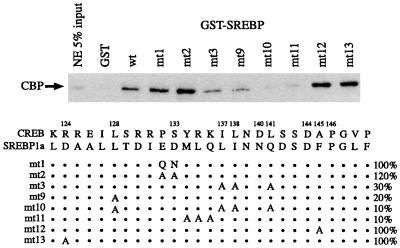
FIG. 2.
Binding of full-length CBP to GST-SREBP mutants. The schematic depicts mutants analyzed at the top. Numbers refer to amino acid positions in CREB; dots refer to retained SREBP residues. (For example, in mt1, the ED in SREBP is replaced by QN.) Percent binding (compared to wild-type SREBP) is indicated on the right. NE, nuclear extract.

