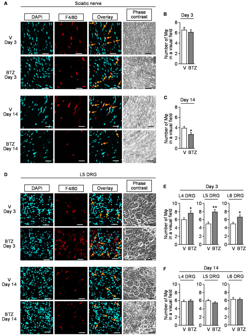
Figure 5.
Immunofluorescence detection of macrophages in the sciatic nerve or dorsal root ganglion of the mice with CIPN caused by bortezomib. Bortezomib at 0.4 mg/kg or vehicle was administered i.p. on days 0, 2, 5, 7, 9, and 12. The sciatic nerves (A–C) and dorsal root ganglion (D–F) were isolated from the mice on day 3 or 14 after the onset of bortezomib treatment. (A,D) Typical microphotographs for the immunofluorescence staining of F4/80-positive macrophages (arrows) in the sciatic nerve (A) and L5 dorsal root ganglion (D). Blue, DAPI (nucleus); red, F4/80 (macrophage); scale bar, 50 μm. (B,C,E,F) The number of F4/80-positive cells in a visual field of the sciatic nerve (B,C) and L4–L6 dorsal root ganglion (E,F) excised on day 3 (B,E) and day 14 (C,F). Mφ, macrophage; V, vehicle; DRG, dorsal root ganglion; BTZ, bortezomib. Data show the mean with S.E.M for 20 visual fields from 5 mice. * p < 0.05, ** p < 0.01 vs. V.

