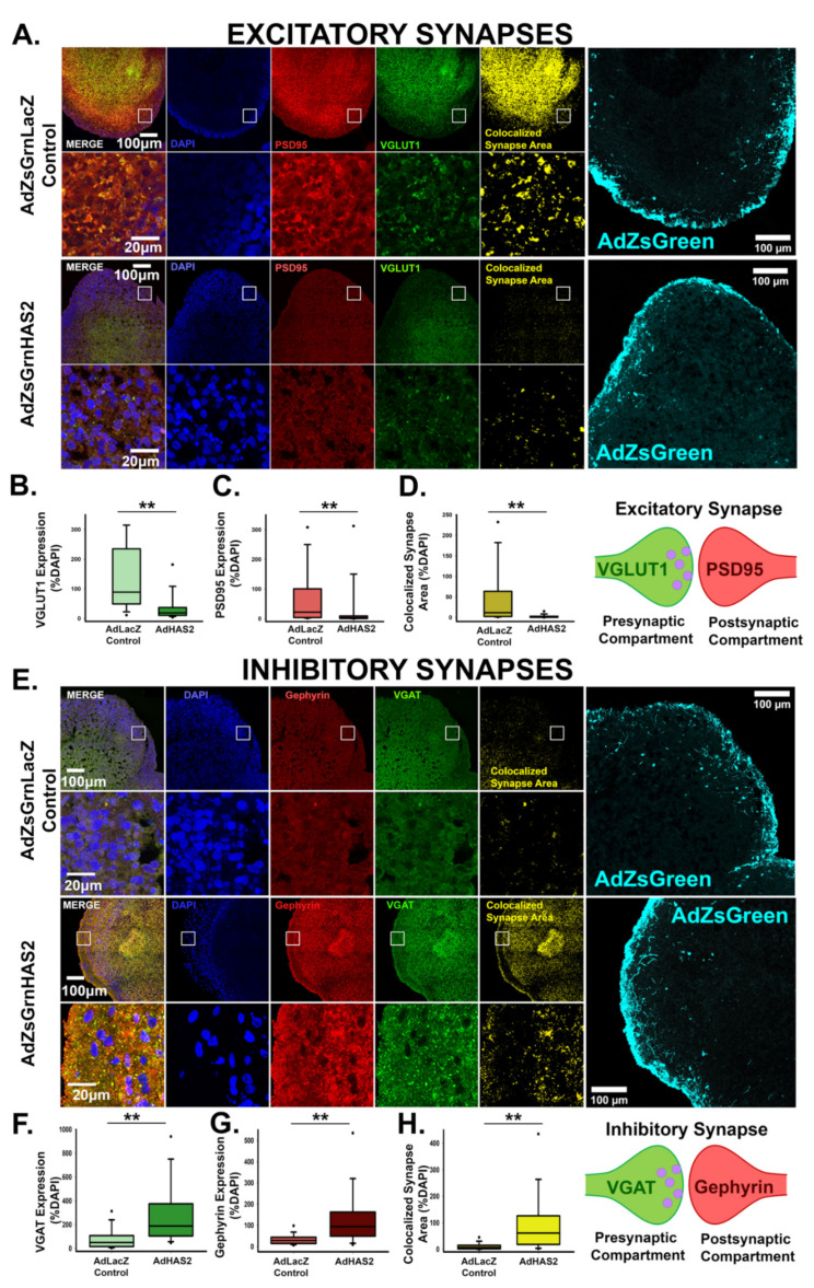
Figure 4.
Hyaluronan synthase, HAS2, alters the emerging balance between excitatory and inhibitory synapses in developing neural networks. (A) Representative images of cortical spheroids transduced with either LacZ control or HAS2 and stained for DAPI (blue) and excitatory synapses: post-synaptic marker PSD95 (red) and pre-synaptic marker VGLUT1 (green). Right panel shows the excitatory synaptic colocalization of PSD95 and VGLUT1 in yellow. Bottom panel highlights the region of the cortical plate indicated by the white box. Scale bars: top: 100 μm, enlarged region: 20 μm. (B) Quantification of the area of pre-synaptic marker VGLUT1 normalized to the area of DAPI. (C) Quantification of area of post-synaptic marker PSD95 normalized to the area of DAPI. (D) Quantification of colocalized excitatory synapse area normalized to the area DAPI. n = 9 total spheroids for each transduction (AdZsGrnLacZ and AdZsGrnHAS2). Spheroids were derived from 3 separately grown and transduced spheroid sets ** p < 0.001 for (B–D). (E) Representative images of transduced cortical spheroids stained for DAPI (blue), and inhibitory synapses: post-synaptic marker gephyrin (red) and pre-synaptic maker VGAT (green). Rightmost panel shows the inhibitory synaptic colocalization of gephyrin and VGAT in yellow. Bottom panel highlights the region of the cortical plate indicated by the white box. Scale bars: top: 100 μm, enlarged region: 20 μm. (F) Quantification of the area of pre- synaptic marker VGAT normalized to the area of DAPI. (G) Quantification of the area of post- synaptic marker gephyrin normalized to DAPI. (H) Quantification of colocalized inhibitory synapse area normalized to the area of DAPI. n = 9 total spheroids for each transduction (AdZsGrnLacZ and AdZsGrnHAS2). Spheroids were derived from 3 separately grown and transduced spheroid sets ** p < 0.001 for (F–H).

