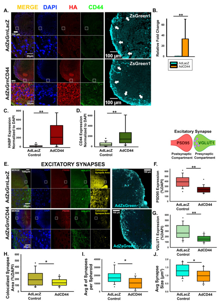
Figure 7.
CD44 antagonizes excitatory synapse formation. (A) Representative images of cortical spheroids transduced with either LacZ control or CD44 and stained for DAPI (blue), HA (red), and CD44 (green). Highlighted region of the cortical plate is enlarged below. Successful transduction was confirmed via viral expression of the fluorophore, ZsGreen1 (cyan, far right panels). Arrows indicate the cortical plate, where synapse analysis was performed. Scale bars top: 100 μm, enlarged region: 20 μm. (B) qRT-PCR confirmation of increased CD44 expression normalized to GAPDH. (C) Quantification of the area of HA (HABP) normalized to the area of DAPI reveals that CD44 promotes HA retention. n = 9 total spheroids for each transduction (AdZsGrnLacZ and AdZsGrnCD44). Spheroids were derived from 3 separately grown and transduced spheroid sets ** p < 0.001. (D) Quantification of the area CD44 expression normalized to the area of DAPI. n = 9 total spheroids for each transduction (AdZsGrnLacZ and AdZsGrnCD44). Spheroids were derived from 3 separately grown and transduced spheroid sets ** p < 0.001. (E) Representative images of cortical spheroids transduced with either LacZ control or CD44 and stained for DAPI (blue), and excitatory synapses: post-synaptic marker PSD95 (red), and pre-synaptic maker VGLUT1 (green). Right panel shows the excitatory synaptic colocalization of PSD95 and VGLUT1 in yellow. Bottom panel highlights the region of the cortical plate indicated by the white box. Successful transduction was confirmed via viral co- expression of the fluorophore, ZsGreen1 (cyan, far right panels). Scale bars: top: 100 μm, enlarged region: 20 μm. (F–H) Quantification of individual pre- and post-synaptic markers and their co-localization in excitatory synapses reveals that CD44 antagonizes excitatory synapse formation. Quantification of the area of post-synaptic marker PSD95 normalized to the area of DAPI. n = 9 total spheroids for each transduction (AdZsGrnLacZ and AdZsGrnCD44). Spheroids were derived from 3 separately grown and transduced spheroid sets. ** p < 0.001 (F). Quantification of the area of the pre-synaptic marker VGLUT1 normalized to the area of DAPI. n = 9 total spheroids for each transduction (AdZsGrnLacZ and AdZsGrnCD44). Spheroids were derived from 3 separately grown and transduced spheroid sets. ** p < 0.001 (G). Quantification of co-localized excitatory synapse area normalized to the area of DAPI. n = 9 total spheroids for each transduction (AdZsGrnLacZ and AdZsGrnCD44). Spheroids were derived from 3 separately grown and transduced spheroid sets. * p = 0.030 (H). (I,J) Quantification of the average number of synapses per spheroid (I) and individual synapse size (J) demonstrates that CD44 promotes the refinement of excitatory synapses into discrete puncta. n = 9 total spheroids for each transduction (AdZsGrnLacZ and AdZsGrnCD44). Spheroids were derived from 3 separately grown and transduced spheroid sets. ** p < 0.001.

