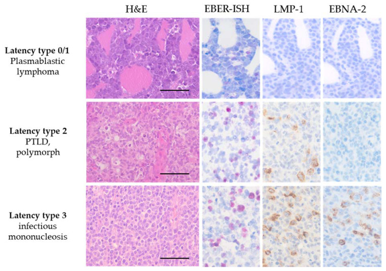
Figure 1.
Micrographs of EBV-associated diseases using histopathological techniques. EBV latency type is correlated with EBV-associated diseases by using EBER-ISH and immunohistochemical stainings with antibodies against LMP-1 and EBNA-2. All latency types show positive signals in the EBER-ISH (pink nuclear signals). Plasmablastic lymphoma, latency type 0 or 1, is negative for LMP-1 or EBNA-2. In contrast, the polymorphic PTLD exhibits positive signals for LMP-1 (brown membranous signal), while immunohistochemistry (IHC) for EBNA-2 remains negative. In infectious mononucleosis, all three markers are positive, and thus a latency type III is determined. H&E stained micrographs show scale bars representing 50 µm.

