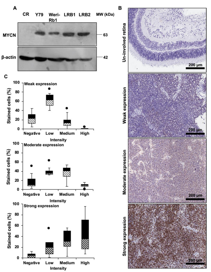
Figure 1.
MYCN is highly expressed in RB. (A) Immunoblot showing overexpression of MYCN in RB cells compared to control retina (CR). (B) Illustrative Immunohistochemical images of MYCN expression in un-involved retina and RB patient specimens showing weak, moderate and strong expression. (C) Box plots depicting the distribution of cells based on intensity scoring of MYCN expression.

