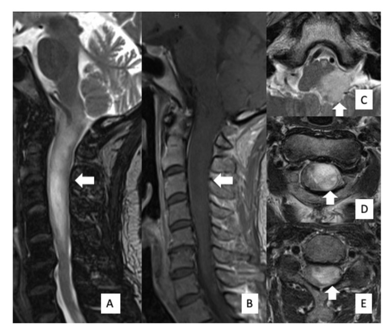
Figure 1.
Preoperative MRI findings. MRI showed an intramedullary mass lesion, extending from the medulla oblongata to C5 (arrow). High signal intensity was evident on T2WI (A) and low signal intensity was evident on T1WI (B). Axial images of T2WI showed that the tumor was disproportionately located on the left side of the spinal cord (C–E).

