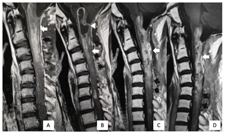
Figure 6.
Postoperative MRI findings. Gadolinium-enhanced T1WI imaging 1 week after surgery revealed a residual tumor (arrow). (A) Two months after the first surgery, cystic lesions appeared from the medulla oblongata to C2 (arrowhead). (B) After syringosubarachnoid shunt for the expanding cyst and tumor re-excision, the cystic lesion disappeared, but the residual tumor was still present (arrow). (C) One year after the first surgery, there was no further regrowth of the residual tumor (arrow) (D).

