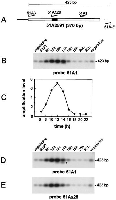
FIG. 1.
Excision of IES 51A2591 and of the internal 28-bp IES in synchronized exconjugant cells. (A) Schematic diagram of IES 51A2591, with the internal 28-bp IES indicated by a solid box. The oligonucleotides used in the study are represented by arrowheads. (B) Southern blot analysis of the PCR products amplified from synchronized exconjugant cells with primers 51A3 and 51A-3′. Samples were run on a 1.5% agarose gel and revealed with 32P-labeled oligonucleotide 51A1. Indicated time points refer to the time following the mixing of reactive cells. Two independent samples of 10 vegetative cells were used as controls for the micronuclear signal. (C) Quantitative analysis of the excision timing of IES 51A2591. The signals shown in panel B were quantified and normalized relative to the value obtained for the 6 h 30 min time point. (D) Prolonged electrophoresis of the samples shown in panel B on a 1.5% agarose gel. IES 51A2591-derived fragments were revealed by Southern blot hybridization with oligonucleotide 51A1. The 400-bp band lacking the 28-bp internal IES is marked by an asterisk. (E) Same blot as in panel D, but hybridized with oligonucleotide 51AΔ28.

