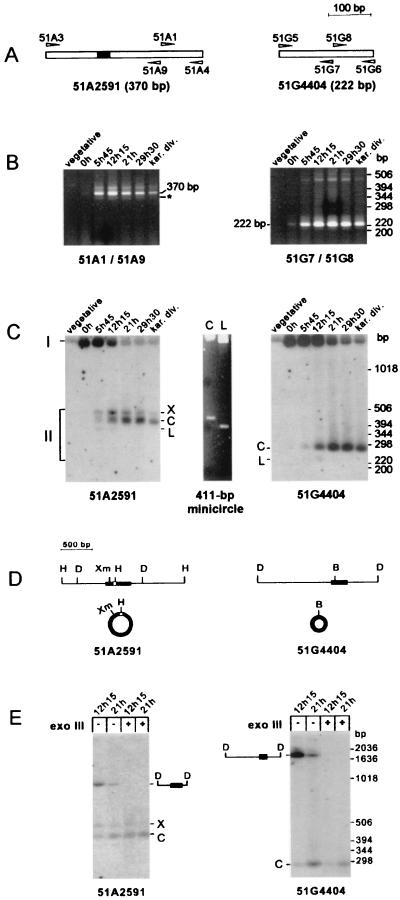
FIG. 4.
Direct detection of circular forms of the excised IESs in autogamous cells. (A) Diagram of IESs 51A2591 and 51G4404, with the corresponding oligonucleotide primers represented by arrowheads. (B) PCR amplification of circular junctions from autogamous cells. Primer sets 51A1 plus 51A9 and 51G7 plus 51G8 were used for the detection of a junction between the ends of IES 51A2591 (left panel) and IES 51G4404 (right panel), respectively (see Materials and Methods). The t = 0 time point was arbitrarily defined as stated in the text, and other time points refer to later stages of autogamy (see the text for details), up to karyonidal division (kar. div.). Electrophoresis was carried out on a 3% NuSieve gel. The major amplification products are indicated by their sizes, and the 342-bp minor band corresponding to the form lacking the 28-bp IES internal to IES 51A2591 is indicated by an asterisk. (C) Southern blot analysis of uncut genomic DNA from autogamous cells. Electrophoresis was carried out on a 1.5% agarose–1.5% NuSieve gel. Chromosomal (I) and extrachromosomal (II) forms of IES 51A2591 (left panel) were revealed after hybridization with a 370-bp PCR fragment specific for the IES sequence and amplified with primers 51A3 and 51A4. A 236-bp fragment, specific for IES 51G4404 and amplified with primers 51G5 and 51G6, was used as a probe in the right panel. The central panel shows ethidium bromide staining of supercoiled (C) or linear (L) forms of a 411-bp control minicircle (see Materials and Methods), electrophoresed on the same gel. (D) Restriction maps of the micronuclear regions around IES 51A2591 (left) and IES 51G4404 (right). Chromosomal IESs are drawn as black boxes, and the 28-bp IES inside IES 51A2591 is shown as a white square. The corresponding circular IES molecules are not drawn to scale. Restriction enzymes: B, BsaI; D, DdeI; H, HinfI; Xm, XmnI. (E) DdeI and exonuclease III (exo III) treatment of total genomic DNA from 100% autogamous cells. DNA extracted from autogamous cells at t = 12 h 15 min and t = 21 h was treated with DdeI and, where indicated, incubated for 2 h at 37°C with 200 U of exonuclease III. Electrophoresis and hybridization probes were as in panel C. IESs are shown as black boxes on the diagrams of their corresponding micronuclear DdeI fragments.

