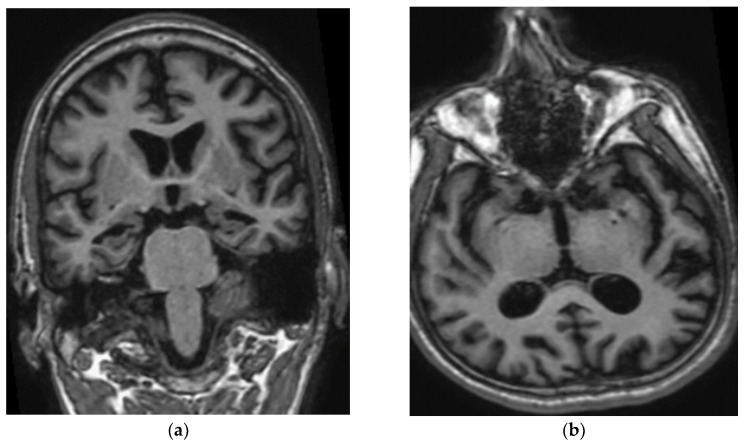
Figure 2.
(a) Coronal T1W MP RAGE image of brain shows atrophy of bilateral hippocampi, more pronounced on the right side. Widening of the cerebral sulci predominantly in the temporal lobes and both lateral ventricles are also noted. (b) Axial T1W MP RAGE image of the brain shows widening of bilateral Sylvian fissure. Dilated occipital horn of both ventricles is also noted.

