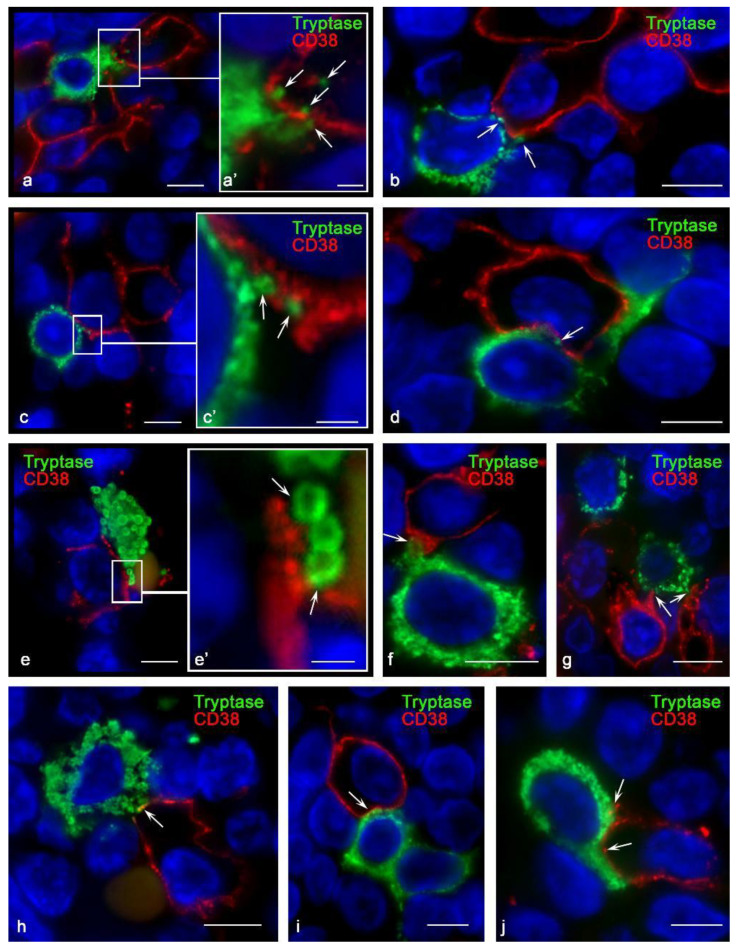
Figure 4.
Histoarchitectonics of specialized immune contact of mast cells with CD38+ cells in tonsilla. Primary antibodies used: (a–e) rabbit monoclonal (Cell Marque, USA); (f–j) mouse monoclonal (kindly provided by Fabio Malavasi, University of Torino, Italy). (a,a′) Mast cell co-localization with multiple CD38+ cells. The enlarged fragment (a′) shows the entry of tryptase-positive granules into the cytoplasm of CD38+ cells towards the nucleus (indicated by the arrow). (b,c,c′,d) Contact of a mast cell with CD38+ cells; various variants of co-localization of tryptase-containing granules with the plasmalemma of CD38+ cells (indicated by an arrow). (e) Interaction of tryptase-positive granules with an exoenzyme on the plasma membrane of a co-localized cell (indicated by an arrow). (f–j) Different morphological variants of the formation of the interaction of mast cell granules with CD38+ tonsillar cells (indicated by an arrow). Scale bar: 1 µm (a′,c′,e′), 5 µm (all others).

