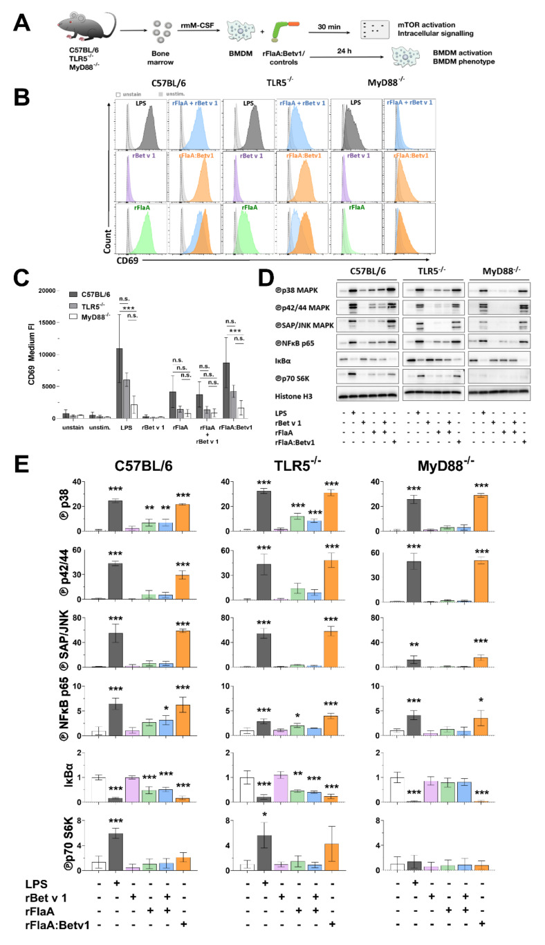
Figure 4.
rFlaA:Betv1-stimulation induces stronger BMDM activation characterized by signaling of MAPK, NFκB, and mTOR . C57BL/6, TLR5−/−, or MyD88−/− BMDMs were differentiated from mouse bone marrow and stimulated with the indicated equimolar protein amounts or LPS as a positive control for either 30 min (Western blot, D) or 24 h (flow cytometry, B,C) (A). Expression levels of CD69 were analyzed by flow cytometry (exemplary result in B, mean fluorescence intensity of three independent experiments depicted in C) and activation of MAPK, NFκB, and mTOR1 signaling was analyzed by Western blot (D). Western blots from three independent experiments were quantified and normalized to expression levels of histone H3 (E). Indicated are statistically significant differences compared to unstimulated controls. Data are either representative results from three independent experiments (B,D) or mean results of three independent experiments ± SD (C,E) with either 10.000 BMDMs (B,C) or one lysate (D,E) measured per experiment. Statistical significance indicated as: n.s. p-value > 0.05, * p-value < 0.05, ** p-value < 0.01, *** p-value < 0.001.

