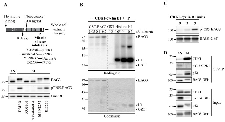
Figure 2.
CDK1 regulates BAG3 phosphorylation dynamic at T285. (A) Schematic of the protocol used. Extracts were prepared from mitotic HeLa-RFP-H2B incubated for 1 h in the presence of chemical inhibitors of the following kinases: RO3306 (CDK1, 8 µM), Purvanolol A (CDKs, 10 µM), MLN8237 (Aurora A, 1 µM), and BI2536 (PLK1, 1 µM) or the vehicle only (DMSO); mitotic cells were collected by a mitotic shake-off. Immunoblotting was performed as indicated; GAPDH: loading control. See also Figure S2. (B) Autoradiogram showing phosphorylation of recombinant GST-BAG3 by a purified CDK1-cyclin B1 complex in the presence of 32 P-ATP; positive control: Histone H1; negative control: GST. (C) Immunoblotting of in vitro phosphorylated BAG3 using the phospho-specific pT285-BAG3 antibody; total levels of GST-BAG3 are shown. (D) BAG3-GFP IPs were prepared from asynchronous (AS) or mitotic HeLa-Flp-In T-REx- BAG3-GFPWT (nocodazole-arrested) transfected with BAG3-specific siRNA (siBAG3 [3′UTR_1], 48 h) and treated with doxycycline to induce BAG3-GFP expression (1 ng/mL, 16 h). Western blots of the GFP IPs were performed as indicated; levels of CDK1, pY15-CDK1 (inactive CDK1), p62, and BAG3-GFP in total cell extracts are shown (Input).

