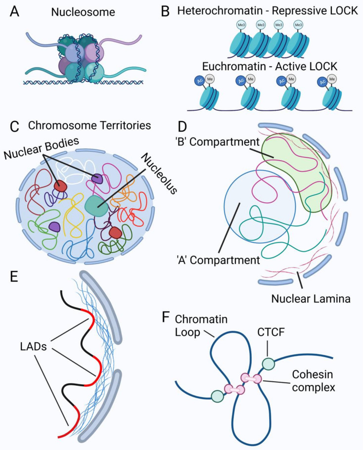
Figure 2.
Chromatin organization in the nucleus. (A) Example of a nucleosome showing DNA wrapped around a histone octamer consisting of two H2A and H2B dimers, two H3 dimers, and two H4 dimers. (B) Example of heterochromatin or closed chromatin (top) and euchromatin or open chromatin (bottom). Long stretches of histones with similar lysine modifications illustrate Large Organized Chromatin Lysine (‘K’) modifications (LOCKs). The closed or open state refers to the accessibility of the chromatin to transcription factors or other DNA binding proteins. (C) Representation of chromosome territories where each colored line depicts one chromosome within the nucleus. Each chromosome is shown occupying its own space within the nucleus. The nucleolus is depicted here interacting with parts of some chromosomes. Nuclear bodies are also present, with examples of Cajal bodies (purple) and PML bodies (red) being shown. (D) Depiction of active ‘A’ compartment in the blue circle, indicating more open chromatin and actively transcribed genes, and ‘B’ compartment, which is mainly heterochromatin and therefore transcriptionally inactive. (E) Examples of Lamina-Associated Domains (LADs), shown here as red chromatin regions. (F) Examples of chromatin loops formed by cohesin complexes and demarcated by CTCF proteins. Created with biorender.com (accessed on 1 September 2021).

