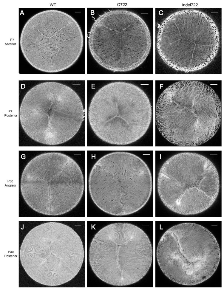
Figure 4.
Whole-mount imaging of Y-suture formation in Epha2-mutant lenses. (A–C,G–I) Representative anterior suture images of wild type (A,G), Epha2-Q722 (B,H), and Epha2-indel722 (C,I) lenses at P7 (A–C) and P30 (G–I). (D–F,J–L) Representative posterior suture images of wild-type (D,J), Epha2-Q722 (E,K), and Epha2-indel722 (F,L) lenses at P7 (D–F) and P30 (J–L). Image depth from lens surface: 100–150 µm (A–L). Scale bar: 100 µm.

